45 label the photomicrogram of the trachea.
quizlet.com › 159345340 › ap-2-lab-unit-2-flash-cardsA&P 2 Lab Unit 2 Flashcards | Quizlet Study with Quizlet and memorize flashcards containing terms like Identify the anatomical structures shown in the anterior view of the superior portion of the lower respiratory system., Put the following layers of the trachea in order from superficial to deep., Label the structures of the upper respiratory system. and more. Trachea (Windpipe): Function and Anatomy - Cleveland Clinic In a diagram of your trachea and other respiratory organs, you can see the trachea between the top lobes of the lungs. It is in front of your esophagus (tube that carries food from your mouth to your stomach). What are the parts of the trachea? The trachea has two parts: Cervical trachea (in your neck). Thoracic trachea (in your chest).
(Get Answer) - Determine the angle of i, r and q this is reflection of ... Determine the angle of i, r and q this is reflection of light General Structure of Mucosa Label the structures that comprise the respiratory tract mucosa (mucous membrane). ... lung Segmental bronchus Trachea prey="" 11="" of="" 46="" next=""> Trachea histology of respiratory system low power Label the photomicrogram of the trachea.

Label the photomicrogram of the trachea.
Unit 6 Flashcards | Quizlet WebLabel the photomicrogram of the trachea. Label the anterior view of the larynx based on the hints if provided. Place the different partial pressure of gases with the appropriate location. Place the following actions with the correct part of pulmonary ventilation. As a result, the alveolar air pressure is _____ the barometric pressure. less than. Inspiration … Trachea (Windpipe) Definition, Anatomy, Function, Diagram Trachea is the medical name for the windpipe, the largest airway in the respiratory system, about 4-5 inches in length and 1 inch in diameter that extends from the lower end of the larynx or voice box [1]. An integral part of the human airway, the trachea, bronchi, bronchioles, and alveoli together make up the lower respiratory tract [2, 3]. Trachea Histology - 4 Layers Identification under Microscope The mucosa of trachea consists of respiratory lining epithelium and lamina propria. In the trachea lining epithelium you will find different types of cells - #1. Pseudostratified ciliated columnar epithelium cells - these are tall, densly packed cell having apical cilia (most predominant in domestic mammals) #2.
Label the photomicrogram of the trachea.. Lung Trachea & Bronchial Tree Diagram & Function | Body Maps - Healthline Bronchial Tree. The trachea, also called the windpipe, is part of the passageway that supplies air to the lungs. Any prolonged blockage, even for a few minutes, can cause death. The trachea is ... Solved Label the photomicrogram of the trachea. Cilia Lamina | Chegg.com Question: Label the photomicrogram of the trachea. Cilia Lamina propria Submucosa Cilia Basement membrane Submucosa Epithelium Basement membrane Lamina propria Epithellum This problem has been solved! See the answer why is it telling me that those are wrong? Show transcribed image text Expert Answer 100% (7 ratings) A & P lab test 4 Flashcards | Quizlet WebLabel the micrograph of the renal corpuscle and surrounding structures using the hints provided. Correctly label the following anatomical parts of a kidney. Classify each of the following parts of the nephron into the correct category based on whether it can only be found in the cortex or if it can be found in the medulla and/or cortex of the kidney. Label The Photomicrograph Of The Lung : 4 Chloro Dl Phenylalanine ... Label the photomicrogram of the lung segmental branch of pulmonary a. Differentiate between trachea, bronchus and bronchiole type i and type ii alveolar cells . A photomicrograph of lung sections from (a) a control group showing normal epithelization of bronchi and bronchioles with normal alveoli and .
quizlet.com › 514038744 › ap-139-chapter-19-flash-cardsA&P 139 Chapter 19 Flashcards | Quizlet Label the photomicrogram of the trachea. Cricoid. Which of these laryngeal cartilages is single? Label these structures of the upper respiratory system. tidal volume. Tracheal Cartilages Anatomy, Function & Diagram | Body Maps - Healthline There are generally sixteen to twenty individual cartilages in the trachea, which varies from person to person. These C-shaped cartilages are stacked one on top of the other and are open at the... label the structures of the upper respiratory system The respiratory system consists of the upper respiratory tract (the nasal cavity, pharynx, larynx, trachea, and bronchi) and the lower respiratory tract (the lungs). As you learn about the various diseases that affect the respiratory system, it is important for you to understand the structures that can be affected by disease. Trachea | Radiology Reference Article | Radiopaedia.org The trachea is a tube-shaped structure consisting of 15-20 D-shaped cartilage rings anterolaterally bridged by annular ligaments. The trachealis muscle (smooth muscle) encircles the trachea completely but is most prominent posteriorly due to the lack of cartilage 4. The trachea extends from the inferior margin of the cricoid cartilage (C6) and ...
APR Respiratory System Flashcards | Quizlet WebLabel the photomicrogram of the trachea. Segmental bronchus and branches. Larynx. Lower Lobe of Right Lung. Cricoid Cartilage. Left Main Bronchus. Frontal Sinus. Students also viewed. A&P 139 Chapter 19. 225 terms. FleetAdmiralJenny. Respiratory System. 24 terms. richtiff12. Unit 6. 354 terms. Nicole_Oliver48. Ch. 23 Assessment . 40 terms. … quizlet.com › 582550133 › apr-respiratory-systemAPR Respiratory System Flashcards | Quizlet Study with Quizlet and memorize flashcards containing terms like Label the structures of the upper respiratory tract., Label the structures of the lower respiratory tract and nearby structures., Assign the following features to the correct anatomical region. and more. Photomicrograph of the trachea Diagram | Quizlet Photomicrograph of the trachea − Learn Test Match Created by monsth3r Plus Terms in this set (8) Cilia ... Pseudostratified ciliated columnar Epithelium ... Seromucous gland ... Hyaline Cartilage ... Collagen Fibers ... Adventitia ... Submucosa ... Mucosa ... Sets found in the same folder LR 3: Heart Auscultation (Define S1 and S2) 2 terms A&P 2 Lab Unit 2 Flashcards | Quizlet WebStudy with Quizlet and memorize flashcards containing terms like Identify the anatomical structures shown in the anterior view of the superior portion of the lower respiratory system., Put the following layers of the trachea in order from superficial to deep., Label the structures of the upper respiratory system. and more.
A&P 2 Lab Unit 2 Flashcards | Quizlet
Trachea - Anatomy & Function - Trachea and Esophagus Location - Health Jade The trachea or "windpipe," is a rigid tube about 12 cm (4.5 in.) long and 2.5 cm (1 in.) in diameter, that lies in front of the esophagus (Figure 1 and 2). The trachea is supported by 16 to 20 C-shaped rings of hyaline cartilage.

Unit 6 Flashcards | Quizlet
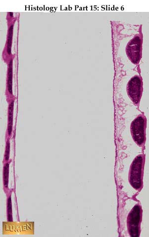
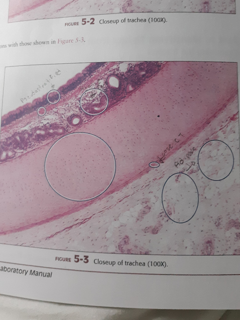
Block1/Fig 4. The microscopic structure of trachea. Fig 4. The microscopic structure of trachea.The lining of the trachea consists of pseudostratified ciliatedcolumnar epithelium. Although the epithelium appears to formthe stratified, but all the cells rest on the basement membrane.The wall of the trachea contains the C-shaped tracheal cartilage.Besides, the tracheal cartilage is surrounded by the regulardense regular connective tissue (DCT).

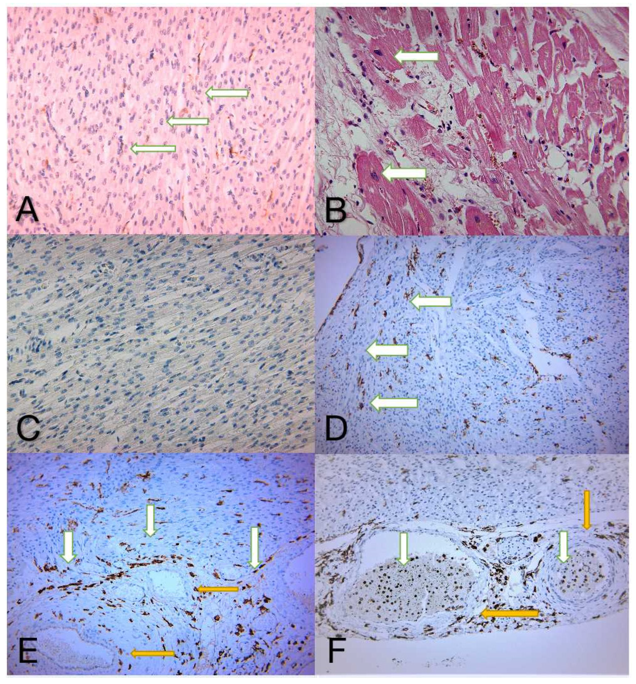
Curcumin and its nano-formulation: The kinetics of tissue ...
Trachea: Definition, anatomy, function, and more - Medical News Today The trachea is a hollow, tube-like structure that runs from the larynx, or voice box, to the bronchi — the two passageways that connect the trachea to the lungs. The average length of the ...
Mammography Education, Inc.
Trachea: anatomy, structure and function - GetBodySmart Many consider the trachea to be the first portion of the lower respiratory tract, which also includes the bronchi, bronchioles, and lungs. The trachea has a wide lumen (= 1 inch or 2.5 cm) and functions to conduct air between the larynx and (primary) bronchi. 1 2 3 Embedded in the wall of the are 16 to 20 tracheal rings made of hyaline cartilage.

Prolonged Inhalation Exposure to Coal Dust on Irradiated Rats ...
Trachea: Anatomy, Function, and Treatment - Verywell Health The trachea, commonly known as the windpipe, is the large tube that delivers air from the upper respiratory tract (the nasal passages, throat, and larynx) to the bronchi (the two large airways that branch off into each lung). In the process, it warms and moisturizes the air and catches debris and microbes before they enter the lungs. 1

Anatomy & Physiology Ch11-Ch14 Flashcards | Quizlet
Label The Photomicrograph Of The Lung : Anatomy Physiology Tissue The ... A photomicrograph of lung sections from (a) a control group showing normal epithelization of bronchi and bronchioles with normal alveoli and . Each individual is unique, so survival rates, treatments and symptoms vary by pati. Light micrograph of lung tissue (click to show / hide labels).

Curcumin and its nano-formulation: The kinetics of tissue ...
Label the photomicrogram of the trachea. - Brainly.com The trachea is known to be a kind of long tube that links the human larynx (voice box) to that of their bronchi. Note that the bronchi is one that send air to a person's lungs and the trachea is known to be an essential part of man's respiratory system. Hence, The Label of the photomicrogram of the trachea is given in the image attached.
Salonul International de Inventica, Bangkok, 2018
The Bronchi: Anatomy, Function, and Treatment - Verywell Health Function. The bronchi function primarily as a passageway for air to travel from the mouth and trachea, down to the alveoli, and back out of the body. 5 In this way, the body's tissues receive oxygen, and carbon dioxide is able to exit the body. Because the bronchi bring in air from outside the body—potentially exposing the lungs to ...

111.pdf - Coronary Sinus Left diagonal artery Right posterior ...
Labeling of the Trachea Quiz - PurposeGames.com Labeling of the Trachea — Quiz Information. This is an online quiz called Labeling of the Trachea. There is a printable worksheet available for download here so you can take the quiz with pen and paper. Quiz Points. 6 p. You need to get 100% to score the 6 points available. Game of the Day. Christmas Song Match EC.

A photomicrograph of a section in rat lung of Group I ...
quizlet.com › 381483065 › a-p-lab-test-4-flash-cardsA & P lab test 4 Flashcards | Quizlet Label the micrograph of the renal corpuscle and surrounding structures using the hints provided. Correctly label the following anatomical parts of a kidney. Classify each of the following parts of the nephron into the correct category based on whether it can only be found in the cortex or if it can be found in the medulla and/or cortex of the ...

Blue Histology - Respiratory System
Histology of trachea and lung - SlideShare 1 of 65 Histology of trachea and lung Mar. 15, 2016 • 104 likes • 79,026 views Download Now Download to read offline Healthcare Histology of trachea and lung mgmcri1234 Follow Advertisement Recommended Histology of respiratory system chanthaj 48.3k views • 42 slides Lecture13 microscopic structure of the respiratory MUBOSScz 14.1k views • 33 slides
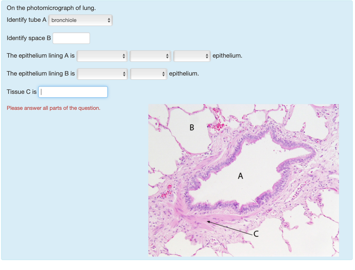
Answered: On the photomicrograph of lung.… | bartleby
quizlet.com › 548952862 › unit-6-flash-cardsUnit 6 Flashcards | Quizlet Label the photomicrogram of the trachea. Label the anterior view of the larynx based on the hints if provided. Place the different partial pressure of gases with the appropriate location.

PDF) English Homophones and Spelling | Andreea Macoviciuc ...
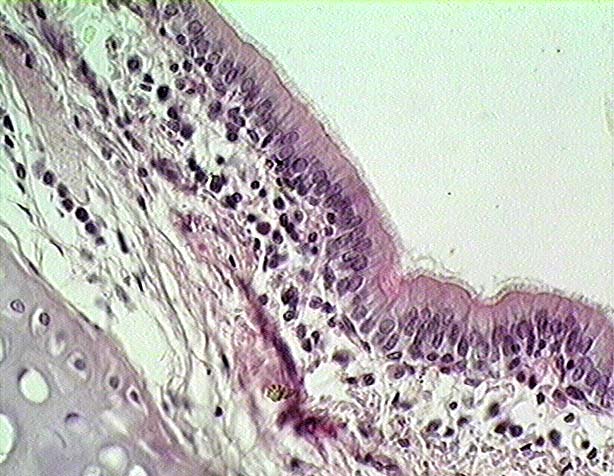
Lab 2: Microscopy and the Study of Tissues - UW-La Crosse The lining of the trachea consists of a type of tissue called pseudostratified (ciliated) columnar epithelium. This single layer of ciliated cells appears stratified because the cells vary in their thickness and because their nuclei are located at different levels. 2 - Pseudostratified columnar epithelium (close-up view) Lab-2 12 Ciliated border

Photomicrograph of the trachea Diagram | Quizlet
Trachea (Windpipe) - Innerbody Trachea (Windpipe) The trachea (or windpipe) is a wide, hollow tube that connects the larynx (or voice box) to the bronchi of the lungs. It is an integral part of the body's airway and has the vital function of providing air flow to and from the lungs for respiration. The trachea begins at the inferior end of the larynx in the base of the neck.
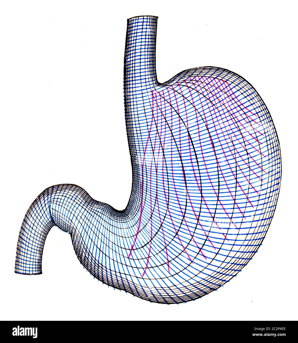
Pyloric hi-res stock photography and images - Page 2 - Alamy
Solved Respiratory Lab Worksheet Saved Help Save & Exit - Chegg Label the photomicrograph of the trachea Show transcribed image text Expert Answer 100% (5 ratings) Transcribed image text: Respiratory Lab Worksheet Saved Help Save & Exit Sbmit Label the photomicrogram of the trachea. Connect 0.23 points Cartilage Print Epithelium References Submucosa Perichondrium Lamina propria Reset Zoom

A & P lab test 4 Flashcards | Quizlet
Picture of the Trachea - WebMD The trachea, commonly known as the windpipe, is a tube about 4 inches long and less than an inch in diameter in most people. The trachea begins just under the larynx (voice box) and runs down...
Slide 107: Trachea
Trachea: Anatomy, blood supply, innervation and function - Kenhub The trachea extends between the larynx and thorax, consisting of two parts; cervical and thoracic. It ends at the level of the sternal angle (T5) where it divides into two main bronchi, one for each lung. Each main bronchus branches out into smaller intrapulmonary bronchi that supply air to the various pulmonary lobes and segments.
Histology Slides 1
A&P 139 Chapter 19 Flashcards | Quizlet WebLabel the photomicrogram of the lung. Label the photomicrogram of the trachea. Cricoid. Which of these laryngeal cartilages is single? Label these structures of the upper respiratory system. tidal volume. The volume of air that enters (or leaves) during a single respiratory cycle is the. Place the following words in order to show the pathway oxygen …
Histologic effects of mandibular protrusion splints in ...
A&P 2 Lab Unit 2 - Quizzes Studymoose Label the structures of the upper respiratory system. answer. question. Match the words on the left with the appropriate definitions on the right. Then put the structures in order from superior to inferior. ... The tracheal cartilage are 13-15 C-shaped cartilage rings.
brave-rewards-ios/wordlist at master · brave/brave-rewards ...
Trachea Histology - 4 Layers Identification under Microscope The mucosa of trachea consists of respiratory lining epithelium and lamina propria. In the trachea lining epithelium you will find different types of cells - #1. Pseudostratified ciliated columnar epithelium cells - these are tall, densly packed cell having apical cilia (most predominant in domestic mammals) #2.
You found the place that will teach you how to find what you ...
Trachea (Windpipe) Definition, Anatomy, Function, Diagram Trachea is the medical name for the windpipe, the largest airway in the respiratory system, about 4-5 inches in length and 1 inch in diameter that extends from the lower end of the larynx or voice box [1]. An integral part of the human airway, the trachea, bronchi, bronchioles, and alveoli together make up the lower respiratory tract [2, 3].
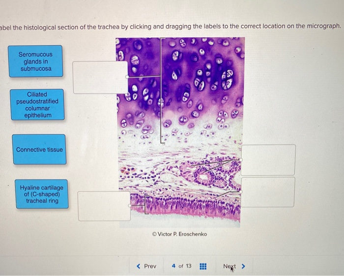
Solved abel the histological section of the trachea by ...
Unit 6 Flashcards | Quizlet WebLabel the photomicrogram of the trachea. Label the anterior view of the larynx based on the hints if provided. Place the different partial pressure of gases with the appropriate location. Place the following actions with the correct part of pulmonary ventilation. As a result, the alveolar air pressure is _____ the barometric pressure. less than. Inspiration …

Curcumin and its nano-formulation: The kinetics of tissue ...
Solved Label this tissue with: Pseudostratified Hyaline ...
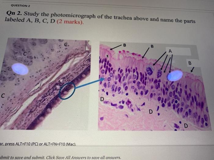
Solved QUESTION 2 On 2. Study the photomicrograph of the ...
14 Technical Aspects of Surgical Neuroangiography
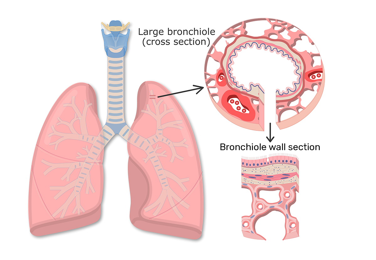
Bronchioles: function and diagram | GetBodySmart

A&P 2 Lab Unit 2 Flashcards | Quizlet
OPTICS & PHOTONICS NEWS APRIL 2019

PDF) English Homophones and Spelling | Andreea Macoviciuc ...

Curcumin and its nano-formulation: The kinetics of tissue ...

Blue Histology - Respiratory System
CASE AT A GLANCE: Partial Epiglottis Removal After Tongue ...

Bronchioles: function and diagram | GetBodySmart

Label the photomicrogram of the trachea. - Brainly.com

A photomicrograph of (a) group I lung tissues showing normal ...

Anatomy & Physiology Ch11-Ch14 Flashcards | Quizlet
Fluorescence In Vivo Endomicroscopy Part 2: Applications of ...
Pyloric hi-res stock photography and images - Page 2 - Alamy
CASE AT A GLANCE: Partial Epiglottis Removal After Tongue ...

111.pdf - Coronary Sinus Left diagonal artery Right posterior ...
Dictionary File | PDF
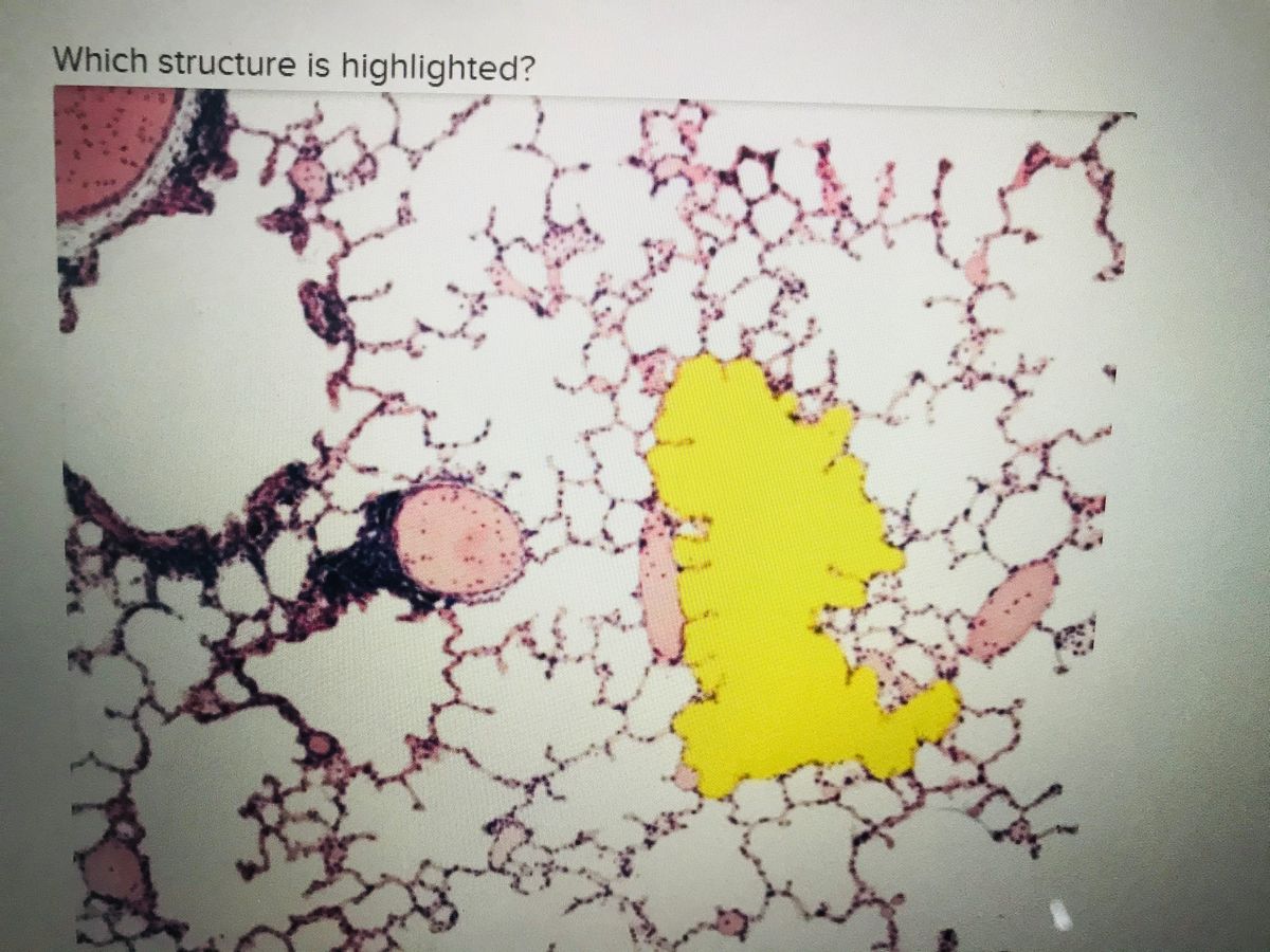
Answered: Which structure is highlighted? | bartleby

Photomicrograph of tracheal wall Diagram | Quizlet

Photomicrograph of trachea of rabbit after 6 weeks ...


Komentar
Posting Komentar