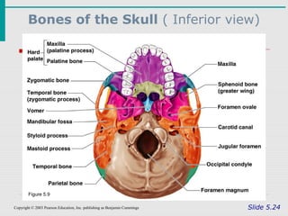
41 inferior skull anatomy labeled
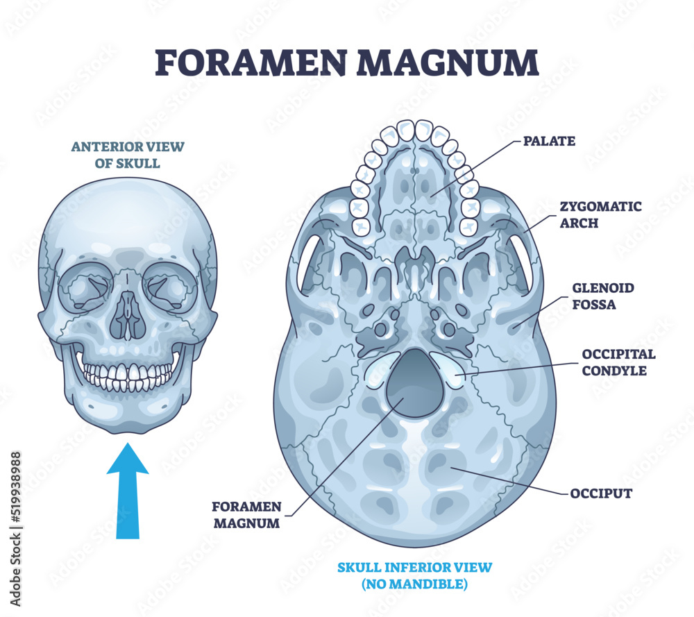
Anatomy, Head and Neck, Suboccipital Muscles - StatPearls - NCBI Bookshelf The suboccipital muscles are a group of four muscles located in the posterior region of the neck, inferior to the occipital bone. These four muscles are the rectus capitis posterior major, rectus capitis posterior minor, obliquus capitis superior, and obliquus capitis inferior. The muscles serve as postural support of the head and neck and allow extension and rotation movements of the neck ... Inferior view of the base of the skull: Anatomy | Kenhub It is an unpaired bone that forms the posterior inferior part of the bony nasal septum. The sphenoid bone sits within the centre of the skull base like a wedge. This bone articulates with the vomer inferiorly, and the greater wings extend laterally to form part of the anterior pterion joint.
PDF Inferior View of Skull (unlabeled) - SLCC Anatomy Inferior View of Skull (unlabeled) Created Date: 3/18/2015 2:45:00 AM ...

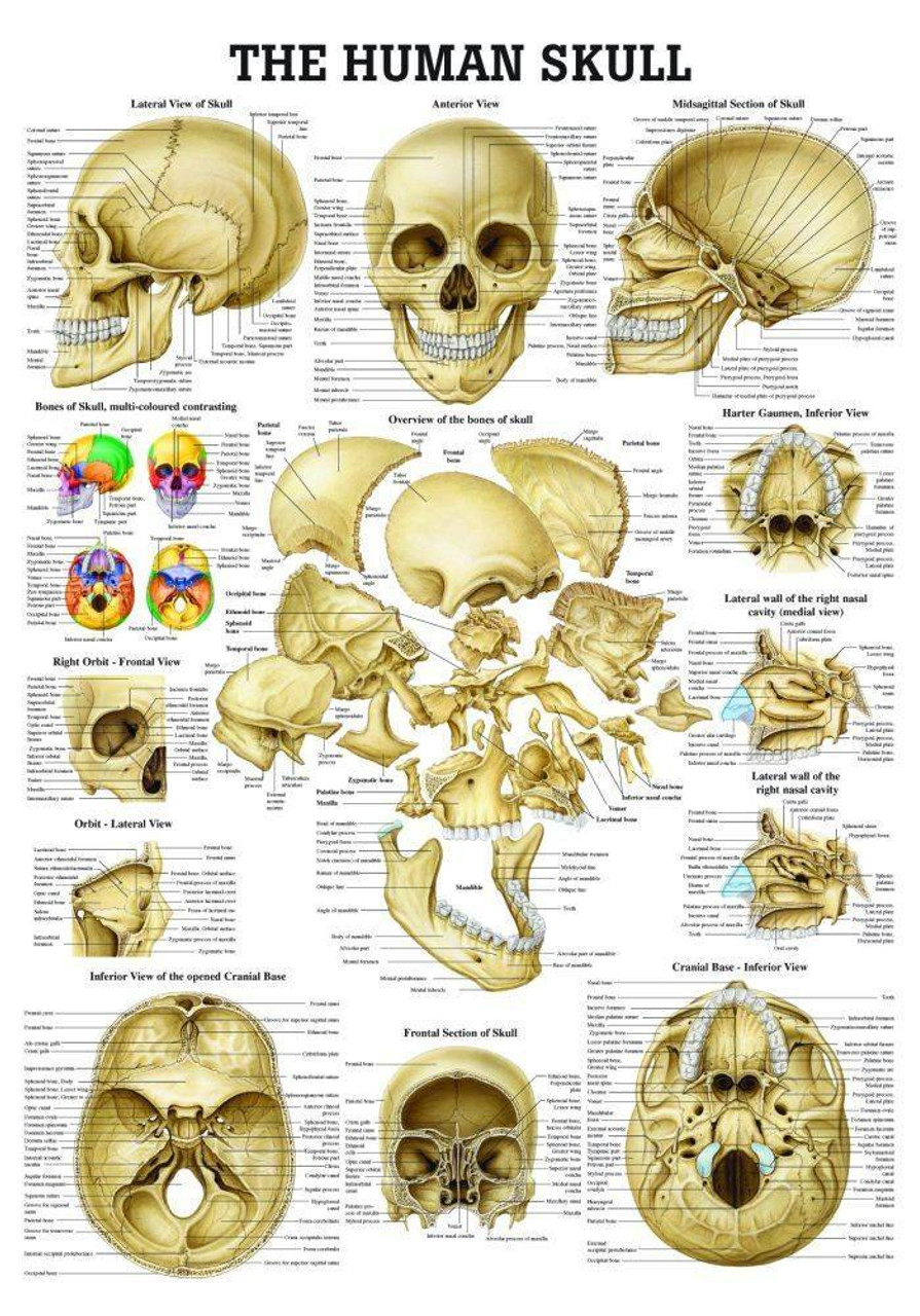
Inferior skull anatomy labeled
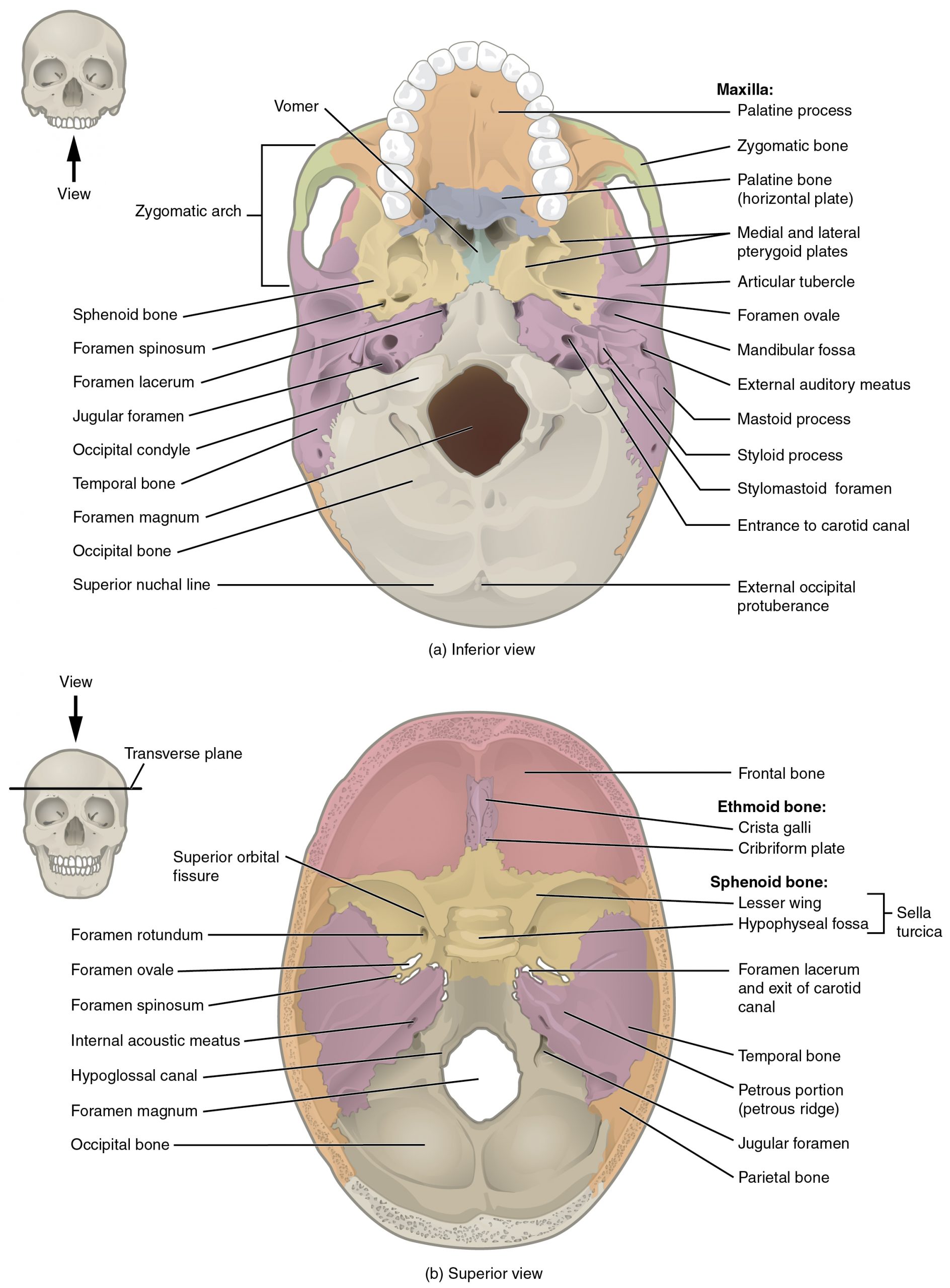
OpenStax AnatPhys fig.7.8 - Superior-Inferior View of Skull Base (b) The complex floor of the cranial cavity is formed by the frontal, ethmoid, sphenoid, temporal, and occipital bones. The lesser wing of the sphenoid bone ... Skull Labeling - Inferior view Flashcards | Quizlet Skull Labeling - Inferior view 5.0 (1 review) zygomatic bone Click the card to flip 👆 Click the card to flip 👆 1 / 15 Flashcards Learn Test Match Created by ryanjmartin98 Terms in this set (15) zygomatic bone sphenoid bone vomer zygomatic process of temporal bone styloid process mastoid process occipital condyle temporal bone C. parietal bone Inferior View of Bony Skull | Neuroanatomy - The Neurosurgical Atlas In this view, the foramen ovale and foramen spinosum can be seen. Posterior to the sphenoid bone in the midline is the clivus, which ends at the anterior margin ...
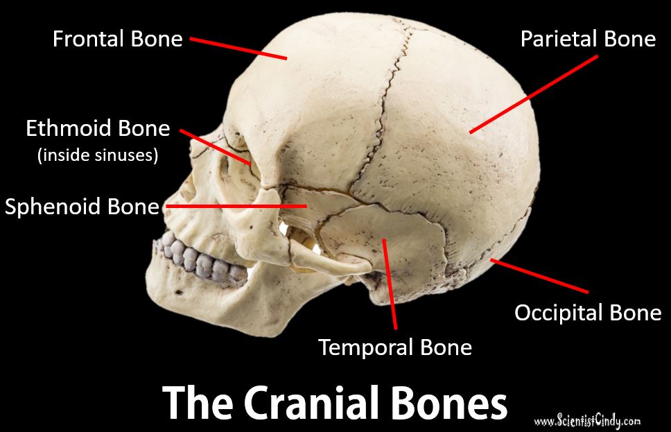
Inferior skull anatomy labeled. Skull | Definition, Anatomy, & Function | Britannica skull, skeletal framework of the head of vertebrates, composed of bones or cartilage, which form a unit that protects the brain and some sense organs. The upper jaw, but not the lower, is part of the skull. The human cranium, the part that contains the brain, is globular and relatively large in comparison with the face. Posterior and lateral views of the skull: Anatomy | Kenhub The inferior border is the zygomatic process laterally and the greater wing of the sphenoid medially. The superior border is demarcated by the two temporal lines that arch across the skull from the zygomatic process of the frontal bone to the supramastoid crest of the temporal bone . Nuchal lines The Skull: Names of Bones in the Head, with Anatomy, & Labeled Diagram Neurocranium. It is the uppermost part of the skull that encircles and protects the brain, as well as the cerebral vasculature and meninges. The hollow space taken up by the brain is called the cranial cavity. The 8 (2 paired and 4 unpaired) bones forming the cranium are called the cranial bones. The cranium is divided into the cranial roof or ... SKELETAL SYSTEM ANATOMY: Inferior aspect of the human skull SKELETAL SYSTEM ANATOMY: Cranial fossa of the human skull · SKELETAL SYSTEM ANATOMY: Mandible · Skull Bones Mnemonic (Cranial and Facial Bones) | ...
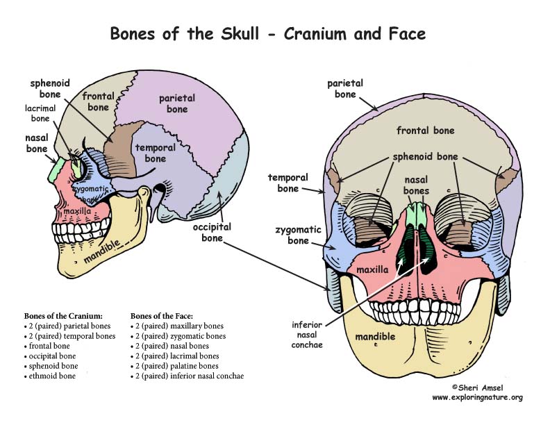
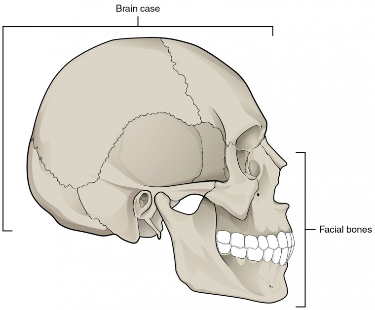
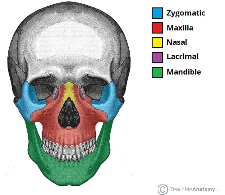
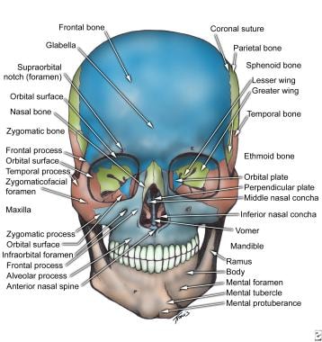
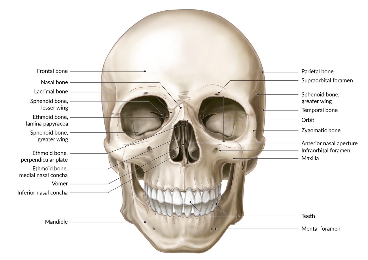
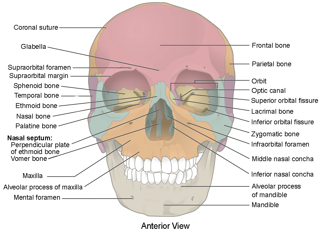
Bones of the Skull - Structure - Fractures - TeachMeAnatomy The cranium (also known as the neurocranium) is formed by the superior aspect of the skull. It encloses and protects the brain, meninges, and cerebral vasculature. Anatomically, the cranium can be subdivided into a roof and a base: Cranial roof - comprised of the frontal, occipital and two parietal bones. It is also known as the calvarium. Skull anatomy: Anterior and lateral views of the skull | Kenhub The human skull consists of about 22 to 30 single bones which are mostly connected together by ossified joints, so called sutures. The skull is divided into the braincase ( cerebral cranium) and the face ( visceral cranium ). The main task of the skull is the protection of the most important organ in the human body: the brain. Human Skeleton Anatomy - Pinterest Skull labeled Human Skeleton Anatomy, Human Anatomy Drawing, Anatomy Bones, ... 1) Frontal processs of maxilla (Figures 1 & 6) Inferior orbital fissure (Fi… 10.3: The Skull - Biology LibreTexts When looking into the nasal cavity from the front of the skull, two bony plates are seen projecting from each lateral wall. The larger of these is the inferior nasal concha, an independent bone of the skull. Located just above the inferior concha is the middle nasal concha, which is part of the ethmoid bone.
Learn skull anatomy with skull bones quizzes and diagrams Skull anatomy diagrams As mentioned, the skull is home to so many structures that the prospect of learning them all can seem very overwhelming. An easy step-by-step system for breaking the topic down then, is essential. For this, we love labeled diagrams. Labeled Skull Diagram Overview image of an anterior view of the skull 7.2 The Skull - Anatomy and Physiology 2e | OpenStax On the inferior aspect of the skull, each half of the sphenoid bone forms two thin, vertically oriented bony plates. These are the medial pterygoid plate and lateral pterygoid plate (pterygoid = "wing-shaped"). The right and left medial pterygoid plates form the posterior, lateral walls of the nasal cavity. The Skull – Anatomy & Physiology - UH Pressbooks When looking into the nasal cavity from the front of the skull, two bony plates are seen projecting from each lateral wall. The larger of these is the inferior ... Inferior View of the Skull - Anatomical Justice This exhibit depicts the anatomy of the inferior skull including: the foramen magnum, occipital condyles, mastoid process, styloid process, mandibular fossa ...
The Skull Bones Anatomy - Inferior View | GetBodySmart Let's start with taking a look at the cranial and facial bones from an anterior view before we dive into their markings from an inferior perspective. Facial Bones: Zygomatic bone ( os zygomaticum ). Maxilla bone ( os maxilla ). Palatine bone ( os palatinum ). Learn skull anatomy faster with these interactive skull bones quizzes and worksheets.
Facial Bones of the Skull Mnemonic: Anatomy and Labeled Diagram - EZmed Nine = Nasal (2) Very = Vomer. Large = Lacrimal (2) Zucchini = Zygomatic (2) Pizzas = Palatine (2) The (2) denotes a pair, or 2, of those bones in the skull. We are now going to discuss the anatomy and important features of each facial bone in the order of the mnemonic. Image: The above mnemonic will help you remember the names of the facial bones.
Skull inferior view Quiz - PurposeGames.com Skull inferior view by ellsanatomy 66,201 plays 19 questions ~50 sec English 19p 111 4.62 (you: not rated) Tries Unlimited [?] Last Played March 18, 2023 - 11:31 PM There is a printable worksheet available for download here so you can take the quiz with pen and paper. From the quiz author Human skull review Remaining 0 Correct 0 Wrong 0 Press play!
The Skull | Anatomy and Physiology I - Lumen Learning On the inferior aspect of the skull, each half of the sphenoid bone forms two thin, vertically oriented bony plates. These are the medial pterygoid plate and lateral pterygoid plate (pterygoid = "wing-shaped"). The right and left medial pterygoid plates form the posterior, lateral walls of the nasal cavity.
Skull: Anatomy, structure, bones, quizzes | Kenhub The skull base is the inferior portion of the neurocranium. Looking at it from the inside it can be subdivided into the anterior, middle and posterior cranial fossae. The skull base comprises parts of the frontal, ethmoid, sphenoid, occipital and temporal bones.
8.2.3: Markings of the Cranium - Biology LibreTexts creates a bridge-like structure that connects the temporal bone with the zygomatic bone forming part of the zygomatic arch. Above: Markings of the cranium with the following views: (A) anterior view, (B) lateral view of the left side of the skull, (C) posterior view, and (D) lateral view of the right side of the skull.
7.3 The Skull - Anatomy & Physiology On the inferior aspect of the skull, each half of the sphenoid bone forms two thin, vertically oriented bony plates. These are the medial pterygoid plate and lateral pterygoid plate (pterygoid = "wing-shaped"). The right and left medial pterygoid plates form the posterior, lateral walls of the nasal cavity.
Bones of the head: Skull anatomy | Kenhub The term 'the skull' includes all the bones of the head, face and jaws. Within this capacity, there are twenty eight individual bones. Of these twenty eight bones, eleven of them are paired, to form a bilaterally symmetrical three dimensional structure and six of them are single, unique bones.
Inferior View of Bony Skull | Neuroanatomy - The Neurosurgical Atlas In this view, the foramen ovale and foramen spinosum can be seen. Posterior to the sphenoid bone in the midline is the clivus, which ends at the anterior margin ...
Skull Labeling - Inferior view Flashcards | Quizlet Skull Labeling - Inferior view 5.0 (1 review) zygomatic bone Click the card to flip 👆 Click the card to flip 👆 1 / 15 Flashcards Learn Test Match Created by ryanjmartin98 Terms in this set (15) zygomatic bone sphenoid bone vomer zygomatic process of temporal bone styloid process mastoid process occipital condyle temporal bone C. parietal bone
OpenStax AnatPhys fig.7.8 - Superior-Inferior View of Skull Base (b) The complex floor of the cranial cavity is formed by the frontal, ethmoid, sphenoid, temporal, and occipital bones. The lesser wing of the sphenoid bone ...

![Human Skull structure - Stock Illustration [6161121] - PIXTA](https://t.pimg.jp/006/161/121/1/6161121.jpg)













Komentar
Posting Komentar