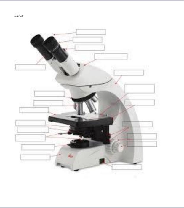
44 label parts of the microscope
Virtual Microscope - NCBioNetwork.org Lesson Description BioNetwork’s Virtual Microscope is the first fully interactive 3D scope - it’s a great practice tool to prepare you for working in a science lab. Explore topics on usage, care, terminology and then interact with a fully functional, virtual microscope. When you are ready, challenge your knowledge in the testing section to see what you have learned. Microscope Imaging Software | Products | Leica Microsystems 23.08.2021 · A range of specific modules allows configuration of the microscope as a dedicated high-performance tool for almost any application. The latest software platform, LAS X, encompasses all microscope solutions for Life Science and Industry applications, offering maximum flexibility. The previous Leica Application Suite continues to be supported.
Virtual Labs: Using the Microscope - GameUp - BrainPOP. In this free online science interactive, students learn the procedures for operating a compound optical light microscope as they would use in a science lab. bVX0-zncj9qJ3G1_r18rkIpQL02X-Oi6tWViR4g4-vwDVmU50WZA-4bRZMjM2TXmc88PAkJ1g0jIembnEbM

Label parts of the microscope
Electron microscope - Wikipedia An electron microscope is a microscope that uses a beam of accelerated electrons as a source of illumination. As the wavelength of an electron can be up to 100,000 times shorter than that of visible light photons , electron microscopes have a higher resolving power than light microscopes and can reveal the structure of smaller objects. Microscope slide - Wikipedia A microscope slide is a thin flat piece of glass, typically 75 by 26 mm (3 by 1 inches) and about 1 mm thick, used to hold objects for examination under a microscope.Typically the object is mounted (secured) on the slide, and then both are inserted together in the microscope for viewing. This arrangement allows several slide-mounted objects to be quickly inserted and … Label the Model Human Cell Quiz - PurposeGames.com This is an online quiz called Label the Model Human Cell . There is a printable worksheet available for download here so you can take the quiz with pen and paper. Your Skills & Rank. Total Points. 0. Get started! Today's Rank--0. Today 's Points. One of us! Game Points. 12. You need to get 100% to score the 12 points available. Actions. Add to favorites 24 favs. Add to Playlist 7 playlists ...
Label parts of the microscope. Label the microscope — Science Learning Hub All microscopes share features in common. In this interactive, you can label the different parts of a microscope. Use this with the Microscope parts activity to help students identify and label the main parts of a microscope and then describe their functions. Drag and drop the text labels onto the microscope diagram. If you want to redo an ... Microscope Parts Quiz - PurposeGames.com Nov 04, 2009 · This is an online quiz called Microscope Parts. There is a printable worksheet available for download here so you can take the quiz with pen and paper. Your Skills ... Parts of a microscope with functions and labeled diagram 17.09.2022 · Thank you very much it really helped me with my science home work since i in 8th grade and this my home work to draw a microscope label all the parts and the function thank may the holy father of holy spirits bless you and give more wisdom thanks love you all keep up the good work and thank you again bye. Reply . Sagar Aryal. May 21, 2022 at 1:57 AM . Thank you … Labeling the Parts of the Microscope Labeling the Parts of the Microscope. This activity has been designed for use in homes and schools. Each microscope layout (both blank and the version with answers) are available as PDF downloads.
Microscopy Pre-lab Activities - University of Delaware Microscope controls: turn knobs (click and hold on upper or lower portion of knob) throw switches (click and drag) turn dials (click and drag) move levers (click and drag) changes lenses (click and drag on objective housing) select a specimen (click on a slide) LabRAM HR Evolution - HORIBA Confocal Raman Microscope. The LabRAM HR Evolution Raman microscopes are ideally suited for both micro and macro measurements, and offer advanced confocal imaging capabilities in 2D and 3D. The true confocal Raman microscope enables the most detailed images and analyses to be obtained with speed and confidence. With guaranteed high performance ... Parts of the Microscope with Labeling (also Free Printouts) Mar 07, 2022 · Click to Download : Label the Parts of the Microscope with answers (A4) PDF print version. For a thorough review of each microscope part continue reading…. A basic microscope has a single convex lens such as those found in a magnifying glass, which you can use to visualize the finest prints. Parts of Stereo Microscope (Dissecting microscope) – labeled … Optical parts of a stereo microscope work together to magnify and produce a 3-D image of the specimens. These parts include: Eyepieces. The eyepiece (or ocular lens) is the lens part at the top of a microscope that the viewer looks through. Typically, standard eyepieces for a dissecting microscope have a magnifying power of 10x. Optional eyepieces of varying powers are …
Microscope Labeling Game - PurposeGames.com About this Quiz. This is an online quiz called Microscope Labeling Game. There is a printable worksheet available for download here so you can take the quiz with pen and paper.. This quiz has tags. Click on the tags below to find other quizzes on the same subject. UD Virtual Compound Microscope - University of Delaware ©University of Delaware. This work is licensed under a Creative Commons Attribution-NonCommercial-NoDerivs 2.5 License.Creative Commons Attribution-NonCommercial-NoDerivs 2.5 License. Label the Model Human Cell Quiz - PurposeGames.com This is an online quiz called Label the Model Human Cell . There is a printable worksheet available for download here so you can take the quiz with pen and paper. Your Skills & Rank. Total Points. 0. Get started! Today's Rank--0. Today 's Points. One of us! Game Points. 12. You need to get 100% to score the 12 points available. Actions. Add to favorites 24 favs. Add to Playlist 7 playlists ... Microscope slide - Wikipedia A microscope slide is a thin flat piece of glass, typically 75 by 26 mm (3 by 1 inches) and about 1 mm thick, used to hold objects for examination under a microscope.Typically the object is mounted (secured) on the slide, and then both are inserted together in the microscope for viewing. This arrangement allows several slide-mounted objects to be quickly inserted and …
Learning Task 3: Label Me!Label The Parts of the Microscope ...
Electron microscope - Wikipedia An electron microscope is a microscope that uses a beam of accelerated electrons as a source of illumination. As the wavelength of an electron can be up to 100,000 times shorter than that of visible light photons , electron microscopes have a higher resolving power than light microscopes and can reveal the structure of smaller objects.

label the parts of microscope scope - Brainly.in

Parts of a Microscope - Free Printable | Free printables ...
Solved Nikon Parts of the compound microscope Write the ...
Science worksheet: Label The Parts Of A Microscope by Science ...

Label the microscope — Science Learning Hub

Parts of a Compound Microscope and Their Functions

Compound Microscope Parts, Functions, and Labeled Diagram ...

Parts of the microscope activity
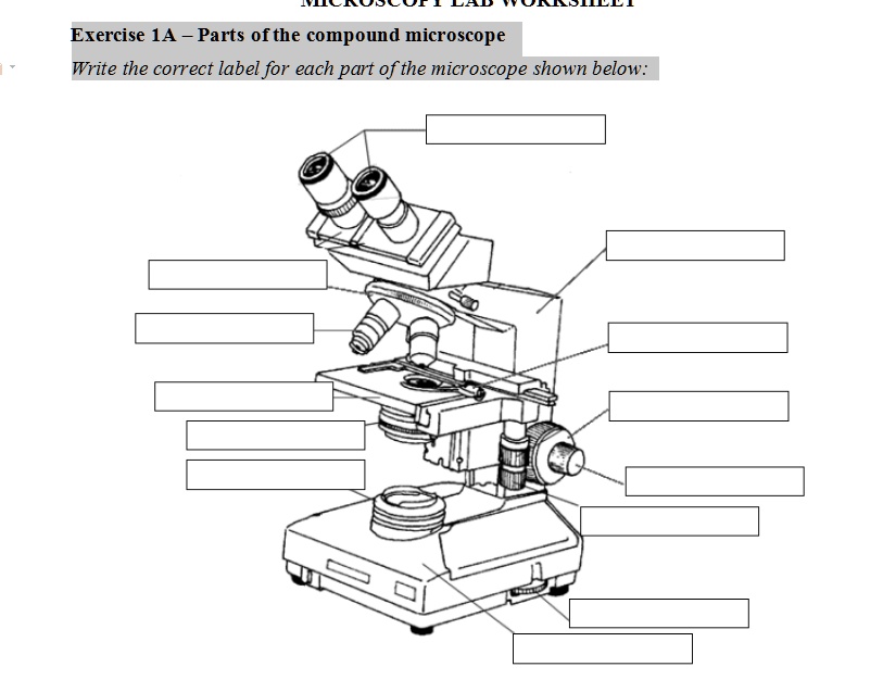
SOLVED: Exercise 1A _ Parts ofthe compound microscope Write ...

Label the Parts of the Microscope - Brainly.ph
7Ac Microscope Labelling Worksheet | Teaching Resources
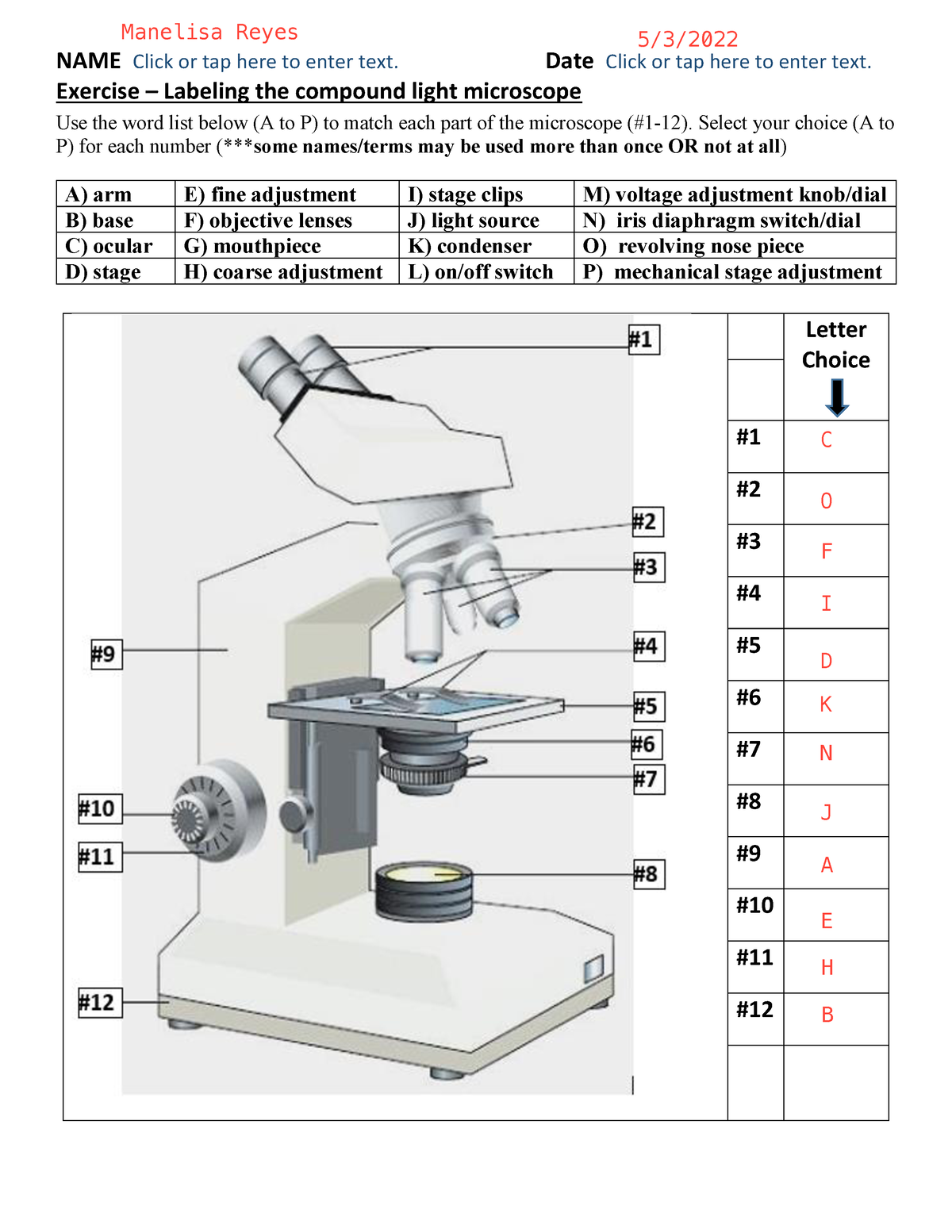
BIO 101 parts of the microscope to label - NAME Click or tap ...
Parts of a Microscope with Their Functions – Microbe Online

Microscope Components - Science Quiz
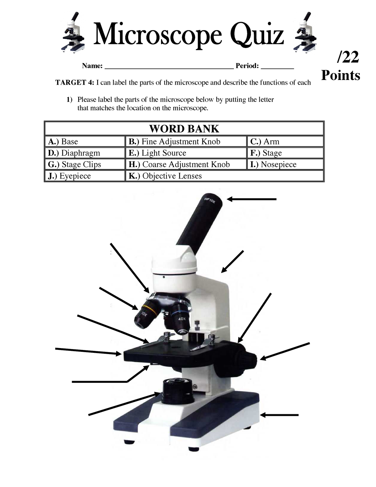
Parts of the Microscope Quiz Lesson 2 - Name: Period: TARGET ...
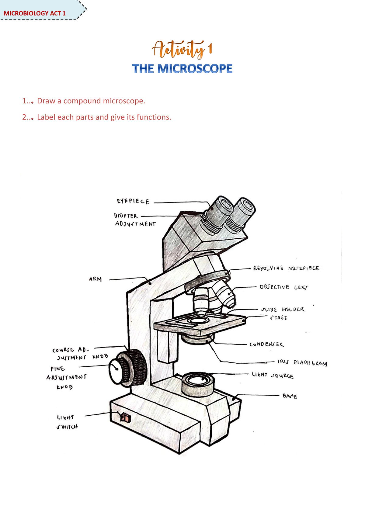
Microscope Activity - MICROBIOLOGY - 1... Draw a compound ...
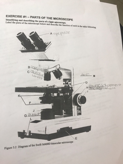
Solved Label the parts of the microscope below and describe ...

Parts of the Microscope worksheet

13 - Microscope Parts - PowerPoint Worksheet.docx - 1 Name: _ ...
Microscope Bundle!! - Parts of a Microscope Unit Activities | TpT

Microscope Maintenance Tips | Science supplies, Multi step ...

Microscope Parts and Functions
Lab - Microscope: MAH-Summer 2019-Anatomy and Physiology I

label microscope diagram | Charts | Microscope, Anatomy bones ...
Compound Microscope. Label the numbered parts of the ...

A Study of the Microscope and its Functions With a Labeled ...

PARTS OF MICROSCOPE| LEARN TO LABEL COMPOUND MICROSCOPE| JUST ...

Dissecting Stereo Microscope Parts and Functions

The Microscope
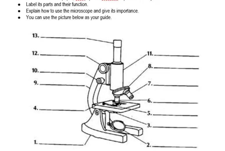
Answered: Label its parts and their function.… | bartleby

Label the microscope — Science Learning Hub

Compound Microscope- Definition, Labeled Diagram, Principle ...

The Parts of a Microscope (Labeled) Printable Printable (6th ...

Compound Microscope Parts – Labeled Diagram and their ...
Name Date Sci STANDARD MICROSCOPE DIAGRAM Label only the ...

Compound Microscope Parts, Functions, and Labeled Diagram ...

Monday 10/19/15 AIM: how do the parts of the compound light ...

Compound Microscope Parts – Labeled Diagram and their ...

Label the numbered parts of the microscope - ppt download

B. ExercisesExercise 1: REMEMBER ME? THEN WRITE MY NAME ...
Label a microscope - Teaching resources
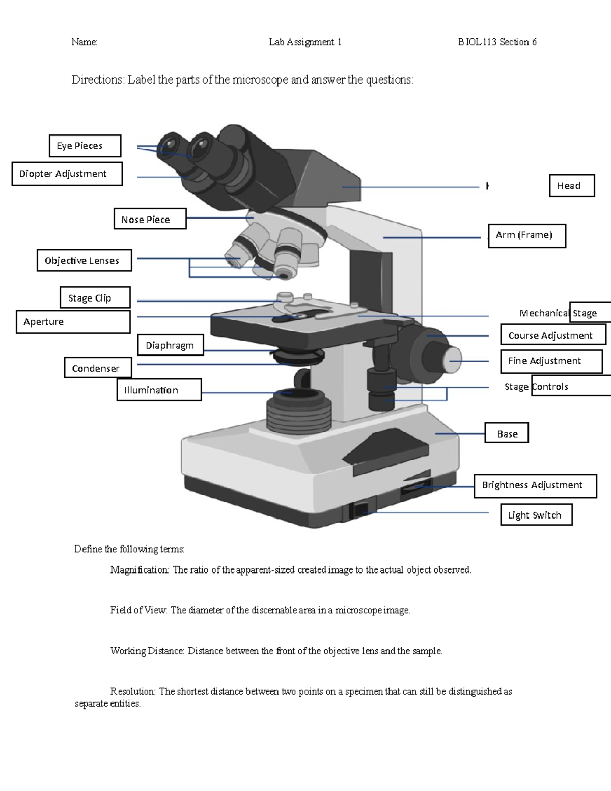
Microscopy Assignment - Name: Lab Assignment 1 BIOL113 ...
Microscope Parts Quiz


Komentar
Posting Komentar